
Visit
Cafmi.Org
For More Summarized Medical Insights & Research
By CAFMI AI From npj Parkinson’s Disease (Open Access)
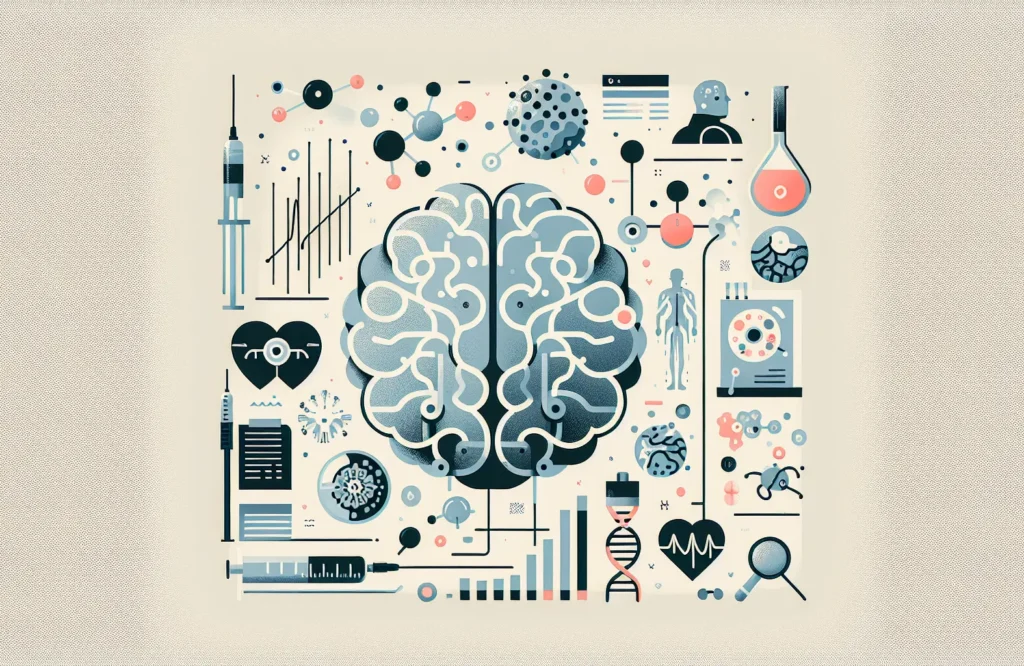
Parkinson’s disease (PD) is a progressive neurodegenerative disorder primarily marked by the abnormal accumulation of α-synuclein (α-syn) proteins in the brain and cerebrospinal fluid (CSF). This aggregation is central to the disease’s pathophysiology and directly influences the neurodegeneration observed in affected patients. Despite significant advances in understanding PD, the ability to detect α-syn aggregation in living patients non-invasively has remained limited, posing challenges in early diagnosis and monitoring of disease progression. The study in question aimed to address this gap by identifying radiological markers that reliably reflect CSF α-syn aggregation, thereby providing surrogate indicators for clinical use. This represents a critical step forward for clinicians, especially those based in the USA, seeking improved tools for managing PD through precise diagnostic and monitoring methodologies.
To achieve this, the research incorporated a cohort of PD patients who underwent comprehensive imaging protocols including magnetic resonance imaging (MRI) and positron emission tomography (PET) alongside lumbar puncture for CSF collection. The CSF samples were analyzed utilizing sensitive biochemical assays specifically designed to quantify α-syn aggregation levels. Parallel to biochemical assessment, imaging data were meticulously processed to isolate brain regions demonstrating significant changes associated with elevated α-syn burden. Key findings revealed strong correlations between increased CSF α-syn aggregation and distinct MRI markers such as reduced brain volume and altered diffusion characteristics predominantly in the substantia nigra and basal ganglia—areas known for their critical role in motor control and widely implicated in PD pathology. PET scans further highlighted abnormal tracer binding in dopaminergic pathways, aligning closely with biochemical markers of α-syn aggregation. The neuroimaging results also paralleled clinical measurements reflecting disease severity and motor dysfunction, thereby supporting the biological relevance and potential utility of these radiological markers in clinical practice.
The identification of specific radiological markers as surrogates for CSF α-syn aggregation carries significant clinical implications. Firstly, these markers offer a promising non-invasive method to evaluate pathological α-syn in patients, potentially enabling earlier and more accurate diagnoses of PD, which is crucial for timely intervention. In practical terms, conventional diagnosis often relies heavily on clinical evaluation and symptom presentation, which may occur after significant neurodegeneration. Radiological detection of α-syn pathology provides a window into the preclinical or early clinical stages, enhancing diagnostic confidence and facilitating stratification of patients for targeted therapies.
Furthermore, these imaging biomarkers have utility in disease monitoring. The study demonstrated that MRI volumetrics and PET tracer binding intensities corresponded with clinical severity, opening possibilities for tracking disease progression quantitatively over time. This is particularly relevant for clinicians involved in longitudinal care and in designing personalized treatment plans. Being able to correlate radiological findings with clinical symptoms enables more precise adjustments of therapeutic approaches and may guide the introduction of novel treatments aimed at α-syn pathology. The non-invasive nature of MRI and PET imaging also supports their practical incorporation into routine clinical workflows, although considerations around cost, availability, and patient suitability remain.
Additionally, the detection of altered diffusion metrics in the substantia nigra and basal ganglia enriches the differential diagnosis framework for movement disorders. Since these brain areas are commonly affected in other parkinsonian syndromes, combining biochemical assays with imaging can help distinguish PD from related conditions. Clinicians can integrate these findings with patient history, clinical examination, and other investigations for more comprehensive assessment. The radiological markers also serve as red flags indicating advancing pathology, prompting timely counseling and discussions related to prognosis, symptom management, and future planning with patients and families.
Looking forward, the integration of radiological markers of CSF α-syn aggregation into clinical practice can transform PD management in several ways. Future research should focus on validating these findings across larger, more diverse populations to ensure generalizability and to refine the sensitivity and specificity of MRI and PET metrics in detecting α-syn pathology. Multi-center studies and longitudinal cohorts would help delineate changes over disease course and determine optimal imaging intervals for monitoring progression.
From a primary care perspective, awareness and education about these emerging diagnostic tools are essential to facilitate timely referrals to specialists and appropriate use of imaging resources. Guidance development involving neurologists, radiologists, and clinicians is necessary to establish standardized imaging protocols, interpretation criteria, and reporting frameworks. Incorporating these radiological markers into existing PD diagnostic criteria or clinical guidelines could enhance diagnostic accuracy and uniformity nationwide.
Patient counseling should also evolve to include discussions about imaging findings, their significance in disease monitoring, and implications for treatment decisions. Clinicians should be prepared to address patient concerns about imaging procedures and ensure shared decision-making regarding follow-up testing. Furthermore, as therapeutic strategies increasingly target α-syn aggregation, radiological markers could play a vital role in selecting candidates for clinical trials and in evaluating treatment efficacy. This comprehensive incorporation of radiological assessment promises to improve outcomes by enabling personalized care pathways and facilitating early intervention in Parkinson’s disease management.
Read The Original Publication Here